
By Rebecca Gibbel MS DVM
WHAT IS FIBROPAPILLOMATOSIS?
Sea turtles are important components of marine ecosystems. They help keep algae in check by their continual grazing, which contributes to the health of coral reefs1. Baby sea turtles face low odds of survival, having only a 1 in 1,000 to 1 in 10,000 chance of surviving to adulthood2 due to predation and pollution. If they are extremely lucky they can live between 50 to 100 years. Unfortunately, since the 1980s, turtles have been increasingly afflicted with a debilitating infectious disease called fibropapillomatosis, or FP3,4. Turtles with this disease develop tumors that usually grow on the head, particularly around and on the eyes. The growths may also be seen on the turtle’s shell and sometimes in the abdomen, where they disrupt organ function and can be fatal. While the tumors are not cancerous, they can grow to significant sizes that interfere with vision, swallowing, breathing, and movement4. The disease develops from a herpes virus and other contributing factors. Genetics and stressors, like environmental pollution and ocean warming, may all play roles in tumor formation.
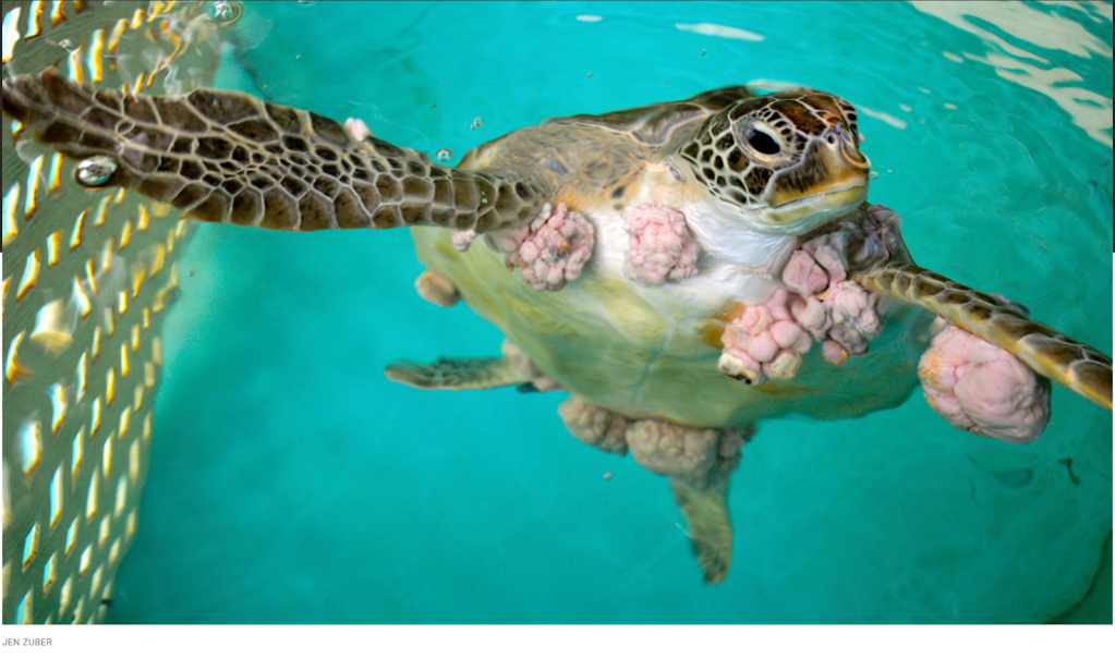
Figure 1. A green turtle with extensive fibropapilloma tumors.
DISEASE BEHAVIOR
Fibropapillomatosis is found worldwide, which is not surprising given the vast distances that sea turtles travel in migration. Up to 70% of sea turtles in Florida and 60% of green sea turtles in Hawaii are now affected with the disease4,3. Green turtles are the most frequently and severely affected, although all 7 species of sea turtles are susceptible. Fibropapillomatosis has been shown to be a transmissible disease and is strongly associated with the virus known as chelonid herpesvirus 5, or ChHV5.
The causality between this herpes virus and the disease is a nuanced one rather than a simple cause and effect. It may be surprising to think of a contagious virus that can cause tumors, but a number of tumor-associated viruses are already known in animals, including humans5. Although turtle hatchlings do not show signs, juveniles are the most frequently affected and may become diseased when they first leave the open ocean and move to coastal areas, where they mingle with other turtles and face multiple detrimental conditions- mostly of human origin. Breaks in the skin surface are probably necessary for virus transmission, and the bites of blood sucking leeches, marine fish, and even contact with other turtles are likely routes of infection.
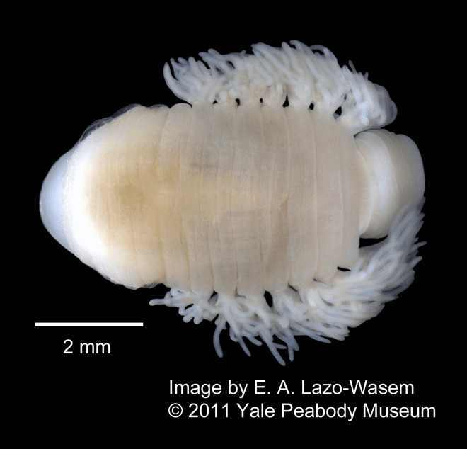
Figure 2. As if a blood sucking Ozobranchus branchiatus leech that preys on green turtles isn’t awful enough,
these nasty parasites have been found to contain the ChHV5 virus.
Older turtles are less likely to be diseased, and it’s not yet known whether they appear spared because the younger turtles have died from the disease and did not live to older ages, or whether the disease regresses as the turtles age, or both. Although they usually grow back, tumors can be surgically removed and even treated with fluoruracil chemotherapy, which is usually not practical in oceanic wild animals. Different species of turtles have differential susceptibility, and there are several strains of the associated virus too. Since the manifestation of the disease varies under different conditions, there seems to be a complex relationship between the microbial agent, the host’s immune response, and the environment.
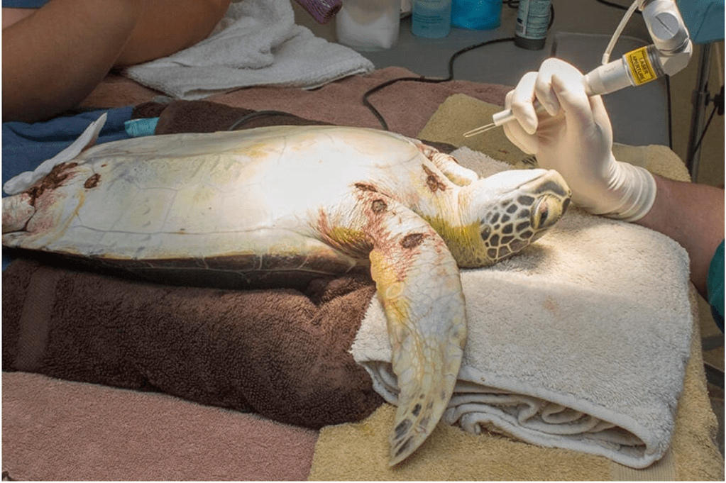
Figure 3. Fibropapilloma tumors being removed with a laser from a sea turtle in rehabilitation.
Photo: Brevard Zoo Healing Center
TIME FOR AN UPDATE?
Koch’s Postulates are the traditional tests for proving that a particular microorganism like a bacterium or virus is the cause of an infectious disease. These criteria were proposed in 1890, before knowledge of viruses. Under these rules6 a suspected pathogen is proven to spread disease a suspected disease pathogen is proven only if all the following conditions are met:
1. The suspected microbe is not present in a healthy host but is found in all diseased hosts
2. The microorganism must be isolated from the diseased organism and grown in laboratory culture media
3. The suspected pathogen must cause the same disease when inoculated into a second susceptible organism
4. The suspected virus, fungus or bacterium must be re-isolated from the second organism
There are numerous reasons why these postulates may not be valid for complex marine diseases. A condition’s cause may be multifactorial, such as diseases caused by microbes in combination with environmental effects, or by more than one microbe acting together. An updated version of Koch’s postulates is needed since not all pathogens grow in the laboratory culture media that were established for terrestrial bacterial organisms- especially marine pathogens. Viruses and fungi are far more difficult to grow in culture, and their identification often relies on creating antibodies to bond with them or by identifying by other means like microscopy or DNA isolation.
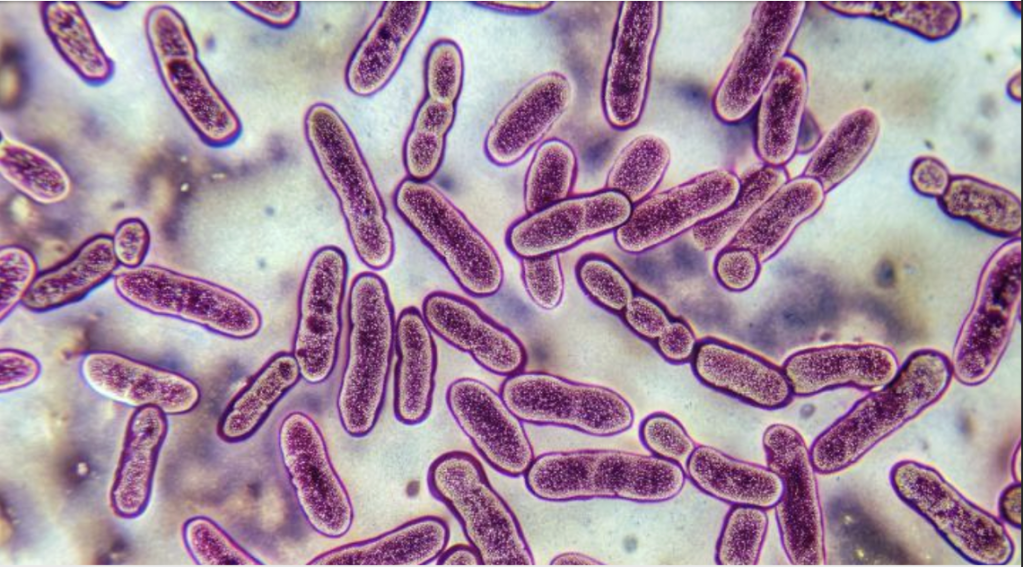
Figure 4: The tuberculosis mycobacterium is an example of a pathogen that does not appear to satisfy Koch’s postulates, despite having been accepted as the cause of the disease for many years. This bacterium takes months to grow in culture, if it grows at all. However, it can be seen readily without cultures simply by examining a sputum or tissue sample under the microscope.
Photo: Institute of Infectious Disease and Molecular Medicine
The list of issues with the Koch postulates continues – another issue is that some wild hosts of disease, like sea turtles and corals are endangered species which are protected and minimally available for testing. It is also difficult to assess whether a host is truly unaffected or if it is subclinically infected without obvious signs. Finally, in this age of environmental degradation, a key point is that an infectious disease may only be manifested if the host is immunocompromised.
An updated version of Koch’s postulates is needed for consideration when a suspected microbe does not satisfy all the criteria. This should allow identification of causality when there is identification of the DNA and/or RNA of the potential pathogen. These revised criteria could be applied to the study of puzzling marine infectious diseases, such as stony coral tissue loss disease and fibropapillomatosis. The newer, more flexible postulates that I’ll informally call Koch+ do establish causality if:
- Nucleic acid sequences (DNA and/or RNA) of the suspected pathogen are present in most cases of the infectious disease and should be found preferentially in the anatomic sites that are affected. Fewer or no sequences should be found in unaffected hosts or tissues.
- If the disease resolves, the number of nucleic sequences of the suspected pathogen should decrease. With relapse of the disease, the opposite should occur, and disease severity should correlate with higher pathogen numbers.
HOW DOES ChHV5 FIT “KOCH +” ?
The ChHV5 virus is consistently found in sea turtles’ fibropapilloma tumors, though it is occasionally also detected in the skin of healthy appearing animals7. Under Koch+, the presence of the alphaherpes viral DNA in the majority of the tumor cells, with healthy cells only very rarely containing those sequences is adequate to demonstrate the causality, though it is important to continue to search for additional environmental influences. The occasional presence of the viral DNA in healthy-appearing turtles suggests that the virus itself may have a latent or dormant state, or that unaffected turtles may be asymptomatic carriers.
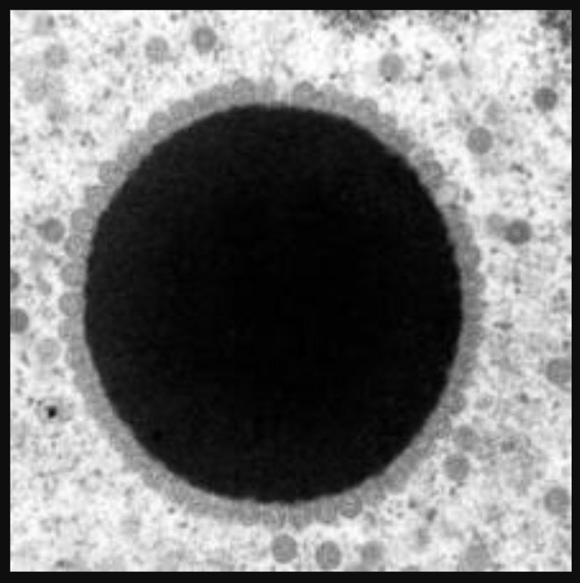
Figure 5. An electron microscopic image of chelonid herpesvirus 5
Photo: usgs.gov/media/images /chelonid-herpesvirus-5-replication
ON THE HUNT FOR COFACTORS
While widespread through oceans, FP has different disease manifestations based on geographic locations4, which argues against a simple “one disease: one pathogen” model. Cases of FP tumors are observed in high numbers in warm coastal areas, but rarely in the open ocean (where it’s hard to observe turtles anyway). It’s thought that viral transmission is high when young turtles leave the pelagic ocean areas and congregate near the shorelines which tend to be areas of high human density with associated pollution from land7. This observation supports two theories- the first is that young turtles become infected when they first encounter a coastal environment with virus in the water, in other turtles, and in parasites. The second hypothesis assumes that most turtles are already carrying the virus, with the fibropapillomatosis disease developing secondary to the additional pressure of degraded water quality closer to shore7.
Intriguingly, papilloma tumors in other species, like the similar-appearing bovine papilloma virus disease, can be potentiated by quercetin, which is a chemical factor in foraged bracken ferns8, which raises the possibility that substances in the turtles’ diet may conceivably play a role. Indeed, Landsberg et al. (1999) observed overlap in high FP areas with the presence of dinoflagellate microalgae on seagrass. The algae contain a tumor promoting compound known as okadaic acid which may accelerate tumor formation when ingested by turtles. The ocean environment also carries numerous pollutant chemicals that are absorbed by the animals’ skin, and any one of them could be a cofactor that influences expression of the disease. Even UV sunlight may accelerate tumor formation and that is a particularly hard factor to avoid! It is notable that elevated levels of dissolved metals such as copper, lead and iron are present in turtles that are heavily affected with FP10. And since the disease has become more common, additional ideas are hypothesized to explain the relatively recent increases in FP, including a new mutation of the herpes virus, and/or immunosuppression of the sea turtles due to the stress of living in an increasingly degraded ocean environment.
NOT A HOPELESS SITUATION
While sea turtle fibromatosis is a profoundly distressing situation, it may be possible to reduce its severity by mitigating detrimental environmental factors. If people are motivated to reduce ocean pollution, particularly in coastal areas, and to prioritize research of fibropapillomatosis cofactors, we can play a positive role in addressing this disease. Sea turtles and other marine animals that are adversely affected by degraded water quality act as sentinels of the contaminants and agents that can harm humans too. If we help them by improving the marine environment, we will help ourselves too.
REFERENCES:
- Eckert, K. L., Bjorndal, K. A., Abreu-Grobois, F. A., & Donnelly, M. (2000). Técnicas de Investigación y Manejo para la Conservación de las Tortugas Marinas. Grupo especialista en tortugas marinas UICN/CSE Publicación, 4.
- https://oceanservice.noaa.gov/news/june15/sea-turtles.html#:~:text=Once%20in%20the%20water%2C%20hatchlings,1%2C000%20to%20one%20in%2010%2C000.
- https://www.fisheries.noaa.gov/national/marine-life-distress/fibropapillomatosis-and-sea-turtles-frequently-asked-questions
- Hargrove, S. A., Work, T. M., Brunson, S., Foley, A. M., Balazs, G. H., Girard, A., … & Murakawa, S. K. (2016). Proceedings of the 2015 international summit on fibropapillomatosis: global status, trends, and population impacts.
- McAloose, D. & Newton, A. L. Wildlife cancer: a conservation perspective. Nature Reviews Cancer 9, 517–526 (2009).
- https://microbenotes.com/kochs-postulates-and-its-limitations/#:~:text=Koch’s%20postulate%20forms%20the%20very,pathogenic%20microbiology%2C%E2%80%9D%20and%20his%20contemporaries
- https://www.woah.org/app/uploads/2021/05/herpesvirus-causing-fibropapillomatosis-in-sea-turtles-infection-with.pdf
- Medeiros-Fonseca, B., Abreu-Silva, A. L., Medeiros, R., Oliveira, P. A., & Gil da Costa, R. M. (2021). Pteridium spp. and bovine papillomavirus: partners in cancer. Frontiers in Veterinary Science, 8, 758720.
- Landsberg, J. H., Balazs, G. H., Steidinger, K. A., Baden, D. G., Work, T. M., & Russell, D. J. (1999). The potential role of natural tumor promoters in marine turtle fibropapillomatosis. Journal of Aquatic Animal Health, 11(3), 199-210.
- da Silva, C. C., Klein, R. D., Barcarolli, I. F., & Bianchini, A. (2016). Metal contamination as a possible etiology of fibropapillomatosis in juvenile female green sea turtles Chelonia mydas from the southern Atlantic Ocean. Aquatic Toxicology, 170, 42-51.
