
By Rebecca Gibbel MS, DVM
Beginning in 2013, the rocky intertidal coastlines of Pacific North America appeared as an invertebrate mass murder scene, with uncountable dismembered corpses of starfish in rapid disintegration. The mortality first began near Olympic National Park in Washington state with the ochre sea star, Pisaster ochraceus, and then spread within months from Mexico to Alaska. For people walking along the normally beautiful shores, it was a grisly sight to witness. First the affected sea stars became lethargic with twisted arms, then their limbs disintegrated and fell off, and finally their bodies began to liquify. This apparently new disease was given the melancholic name “Sea Star Wasting Disease (SSWD), although it had been first reported to science in 1898 with sporadic small outbreaks in the intervening years1.
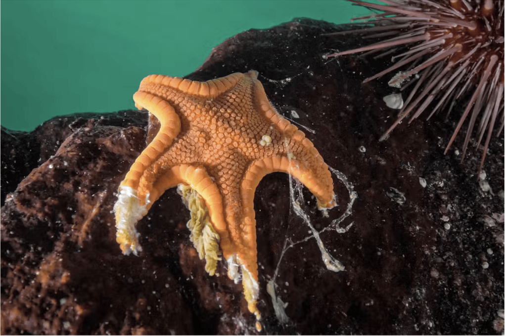
Figure 1. A cookie sea star with SSWD, with disintegrating limbs
Photo credit: Grant Callegari/Hakai Institute
In just five years from the beginning of the mass die off, SSWD caused catastrophic population declines in over 20 species of sea stars, killing an estimated five billion of the most susceptible species, the 24-armed sunflower sea stars (Pycnopodia helianthoide)2. Sea stars play important roles in marine ecosystems and prey up sea urchins, which keeps the urchin populations in check. When the sea star populations became devastated by SSWD, a cascade of disastrous effects resulted. Urchin populations grew massively and they consumed so much kelp that the kelp forests collapsed, along with the enormous numbers of organisms that inhabit them3. In addition to the sea stars’ role as urchin predators, Mancusco et al. (2025) found that the sunflower sea star diffuses chemicals into the surrounding water that elicit “a localized landscape of fear” amongst the urchins, which suppresses their expansion4. This unexpected influence of the sea stars is another biological control that was lost in the SSWD outbreak.
Sea stars are one of the charismatic large faunae that generate affection and concern in humans, and which inspire the creation of cartoon characters like the dumb but happy Patrick Sea Star in the SpongeBob SquarePants show. Captive sea stars will approach researchers and wave their arms about, catching bits of food that are thrown to them. (With 24 arms, they are very good at that trick.)

Fig. 2: Patrick Sea Star character, with all his limbs intact.
Image credit: Wikipedia
Predictably, the mass die-off created great interest and distress among citizens and scientists alike- much more so than with marine animal epidemics, or epizootics, of less lovable creatures like the related spiny sea urchins.
Researchers quickly began searching for the cause of this devastating disease, but for over ten years, answers remained elusive. An important early clue was that sea stars living in nearby aquaria were dying with the same signs. They had no contact with the wild sea stars, but the water for their tanks was drawn from the ocean, which strongly suggested an infectious disease. The tools used in the hunt for answers included microscopic histological examination, bacterial cultures, and genetic profiling of the DNA and RNA of the microorganisms that live within and upon the sea stars.
FIRST CULPRIT
In 2014, Hewson et al. published findings that implicated a densovirus as the cause of SSWD, after that virus was identified in most sick sea stars but not in most of the healthy sea stars that were tested5. The virus was given the name of sea star–associated densovirus (SSaDV) and seemed to be a breakthrough at the time. Although it was initially widely accepted that this virus causes SSWD, subsequent work refuted the finding, with no evidence of a viral cause found with histological examination nor on follow up genetic testing6,7. It may be that virus populations, including SsaDV, are just increased overall in dying sea stars tissue as part of the decay process, rather than a cause of it8. So, the disease detectives resumed their investigations.
BAD ENVIRONMENT?
Next, non-biologic factors were implicated in the onset of the disease. Elevated temperatures, poor water flow, dissolved oxygen depletion, desiccation and abrasion were all found to hasten the wasting condition, both in aquarium experiments and in field surveys9,10,11. But there was still strong evidence of a contagious disease, and these environmental factors alone could not fully explain the apparent transmissibility.
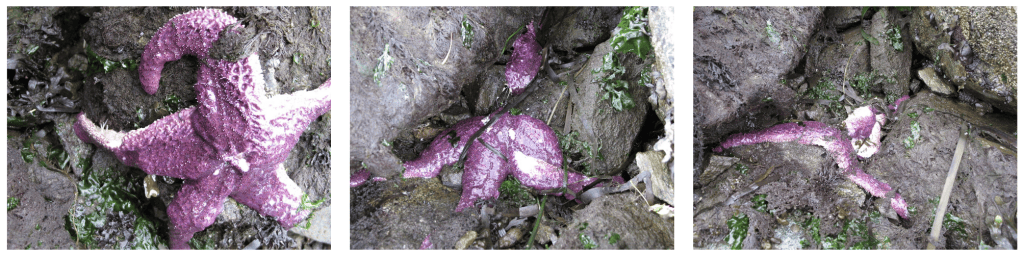
Figure 3. This time series of photographs taken over just three days shows how rapidly SSWD strikes.
Photo: Kit Harma
NEXT: COMPLICATED EFFECTS OF MICROORGANISMS
The next hypothesis that was advanced as a plausible cause of SSWD was the concept that microorganisms at the interface of the sea star animal and the surrounding water consumes oxygen as they break down organic matter (which is a polite description that applies to sewage and rotting material). This leads to oxygen depletion and harm to the sea star11. The theory utilized the field observation that sea stars with rough surfaces die faster of SSWD and hypothesized that as more organic material in the water is trapped on the sea star surface, more bacteria grow in it and remove oxygen. This explanation remained somewhat unsatisfying, since there are always bacteria living on marine organisms and SSWD was established to be a contagious condition, which wasn’t explained by the interface hypothesis. So, the search continued.
Getting closer to The Discovery of the Decade:
In 2023, Grassl et al. published research examining SSWD-infected common sea stars, Asterias ruben, which had originated in the North Sea12. This study performed histopathological light microscopy, bacterial cultures and PCR DNA isolation tests, and found a profound abundance of both Vibrio and Delftia bacterial species. The microscopic examination found intense inflammation which is non-specific as to cause. While suspicious, both of these bacteria were assumed to probably be proliferating because the tissue was decomposing, rather than being the cause of SSWD.
At long last, in 2025 a group of persistent researchers published results from an extensive study that identified the bacterium Vibrio pectenicida strain FHCF-3 as the cause of SSWD in sunflower sea stars (Pycnopodia helianthoides2. The research identified the V. pectenicida bacterium by first sifting through the microbial DNA and RNA of sick versus healthy sea stars with a technique called deep sequencing. First, they identified Vibrio as a bacterial genus that was proliferating in the affected sea stars, and compared its genetic sequence to the 147 known types of Vibrio until the V. pectenicida species was finally identified as the perpetrator. V. pectenidica was seen at highest levels in the visibly abnormal seastars and also found in lower amounts in the exposed but still normal appearing individuals. In starfish from unexposed geographic regions, the bacteria were in very low levels. Once cultures of this bacterium were created, and healthy experimental sea stars succumbed after exposure, the mystery was finally solved.
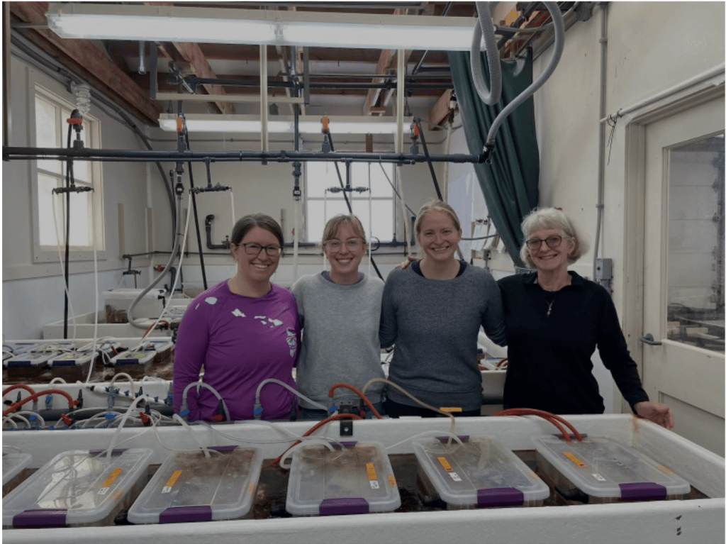
Fig. 4. The team of researchers who finally definitively isolated the causative agent of SSWD, from the USGS Marrowstone Field Station. From left to right: Alyssa Gehman, Grace Crandall, Melanie Prentice and Drew Harvell. Photo credit: Grace Crandall/ University of Washington.
A Sneaky Character:
So how did this bacterium evade detection for over a decade of intensive searching?
Epidemiologists searching for the microbial causes of new diseases have a variety of tools at their disposal, and there is no one technique that works for each investigation. In the case of SSWD, which affects multiple species, the successful marine detectives made a fortuitous choice of starfish species to examine, as well as selecting the right body part to screen.
- The SSWD case was cracked by Deep Sequencing of DNA, which is a tool that has only been available to lab researchers in the past decade. Once DNA sequences are seen in abundance, they must be identified by comparing them to atlases of known organisms. Although the global biodiversity databases are still in development for marine organisms, the V. pectenicida bacterium was identified by comparison to similar sequences from closely related microbes- just as human criminals have been identified by looking at the DNA of relatives.
- The Prentice et al. 2025 study managed to identify the culprit V. pectenicida bacterium because the authors examined the seastars’ “blood” or coelomic fluid. In contrast, the previous histology examinations were done on the solid tissues of the sea stars and failed to find the bacteria there, although the animals’ solid tissues were severely affected.
- Vibrio bacteria need special factors and conditions to allow it to grow in cultures, or they will not be found13. Even different species of Vibrio have different requirements for culture. Once a Vibrio species is known, like human cholera, then specific culture conditions must be used to grow the bacteria.
- Vibrio bacteria can enter a dormant “viable but non-culturable state” which is like an invisibility cloak for bacteria. This state can be induced by environmental stress like starvation, sudden temperature drops, or low salinity14. While the bacteria are alive and still potentially infectious in this state, they will not be found in culture, though their DNA can still be identified.
The Grim Big Picture (sorry):
As our oceans are warming under the effects of climate change, numerous microbial relationships are becoming adversely transformed. Warmer waters create ideal conditions for the proliferation of bacteria, viruses and parasites that affect sea life as well as humans. Vibrio infections in particular, are linked to high mortality in marine organisms as well as significant human health risks15. In marine fish and shellfish, the increasing frequency and severity of Vibrio outbreaks is correlated to rising ocean temperatures which push organisms beyond their stress limits and reduce their ability to fight infections. SSWD is just one more example of the connection between increasing ocean temperatures and marine disease. Vibrio bacteria have been called the barometers of climate change, with billions of seastars now added to the victim list.
REFERENCES
- Mead, A. D. (1898). Twenty-eighth annual report of the commissioners of inland fisheries, made to the General Assembly at its January session, 1898. Southwick JMK, Root HT, Willard CW, Morton WMP, Roberts AD, Bumpus HC (eds).
- Prentice, M.B., Crandall, G.A., Chan, A.M. et al. Vibrio pectenicida strain FHCF-3 is a causative agent of sea star wasting disease. Nat Ecol Evol 9, 1739–1751 (2025). https://doi.org/10.1038/s41559-025-02797-2
- Prentice, J., Gehman, A., Harvell, D., Crandall, G. (2025, August 10). The bacteria killing sea stars in the Pacific: How our team uncovered a decade-long mystery. The Conversation. https://doi.org/10.64628/AAM.7mdyvgv46
- Mancuso, R. T., Gravem, S. A., Campbell, R. S., Hunter, N., Raimondi, P., Galloway, A. W., & Kroeker, K. J. (2025). Sunflower sea star chemical cues locally reduce kelp consumption by eliciting a flee response in red sea urchins. Proceedings of the Royal Society B: Biological Sciences, 292(2050).
- Hewson, I. et al. Densovirus associated with sea-star wasting disease and mass mortality. Proc. Natl Acad. Sci. USA 111, 17278–17283 (2014).
- Bucci C, Francoeur M, McGreal J, Smolowitz R, Zazueta-Novoa V, et al. 2017.. Sea Star Wasting Disease in Asterias forbesi along the Atlantic coast of North America. . PLOS ONE 12::e0188523
- Oulhen N, Byrne M, Duffin P, Gomez-Chiarri M, Hewson I, et al. 2022.. A review of asteroid biology in the context of sea star wasting: possible causes and consequences. . Biol. Bull. 243::50–75
- Hewson, I., Aquino, C. A., & DeRito, C. M. (2020). Virome variation during sea star wasting disease progression in Pisaster ochraceus (Asteroidea, Echinodermata). Viruses, 12(11), 1332.
- Eisenlord, M. E., Groner, M. L., Yoshioka, R. M., Elliott, J., Maynard, J., Fradkin, S., … & Harvell, C. D. (2016). Ochre star mortality during the 2014 wasting disease epizootic: role of population size structure and temperature. Philosophical Transactions of the Royal Society B: Biological Sciences, 371(1689).
- Smith, S., Kunc, H. P., Hewson, I., & Collins, P. C. (2023). Elevated temperature linked to signs associated with sea star wasting disease in a keystone European species, Asterias rubens. Marine Ecology Progress Series, 724.
- Aquino, C. A., Besemer, R. M., DeRito, C. M., Kocian, J., Porter, I. R., Raimondi, P. T., … & Hewson, I. (2021). Evidence that microorganisms at the animal-water interface drive sea star wasting disease. Frontiers in Microbiology, 11, 610009.
- Grassl, K., & Bauer, J. (2023). Sea Star Wasting Disease in captive common sea stars (Asterias rubens): Examinations, therapy attempts and course of disease. Bulletin of the European Association of Fish Pathologists, 43(2).
- Sampaio, A., Silva, V., Poeta, P., & Aonofriesei, F. (2022). Vibrio spp.: Life Strategies, Ecology, and Risks in a Changing Environment. Diversity, 14(2), 97. https://doi.org/10.3390/d14020097
- Tagliavia, M., Salamone, M., Bennici, C., Quatrini, P., & Cuttitta, A. (2019). A modified culture medium for improved isolation of marine vibrios. MicrobiologyOpen, 8(9), e00835.
- Froelich, B. A., & Daines, D. A. (2020). In hot water: effects of climate change on Vibrio–human interactions. Environmental microbiology, 22(10), 4101-4111.

