
Written by Sara Gagliardi
Edited by Danielle Moloney
Culturing cell lines in vitro (i.e., in media cultures at the laboratory) has increased the interest of research. The culture of single-cells provides a system for the study of developmental, regeneration, trans-differentiation, medical, and pharmaceutical biology. Indeed, it is suggested that the culture of coral cells and tissues could reveal important information about coral tissues. First, this experimental system could reveal the cellular and molecular mechanisms of the coral endosymbiosis with dinoflagellate algae from the genus Symbiodinium. These algae are essential to corals. Indeed, they provide the major coral support of energy via photosynthesis. However, Symbiodinium cells are expulsed from coral tissues because of increasing seawater temperatures and diseases, thus endangering coral survival by causing coral bleaching. Therefore, projects aiming at understanding the mechanisms of endosymbiosis could help coral reef sustainability and restoration. Second, coral cell cultures could reveal molecular and cellular mechanisms involved in determining the fates of progenitor cells (or stem cells). Indeed, the body of corals – and Cnidaria – differentiates into two germ layers, ectoderm, and endoderm, with differentiated epithelial cells, neurons, stem cells, a complex extracellular matrix, muscle fibers, and a fixed axis of symmetry. Determining the processes of such differentiation could be achieved with cell culture.
Contrarily to mammal cells, the dissociation of cells and tissues in marine invertebrates is challenging. Tissues from marine invertebrates do not survive in conventional culture medium or in seawater. However, by the end of April 2021, Dr. Kawamura and its colleagues have succeeded in obtaining and maintaining several lines of in vitro cell lines. These cell lines belonged to planula larvae of the coral Acropora tenuis, an acroporid coral found in the Red Sea, the Indian Ocean, and the West and Central Pacific Ocean.
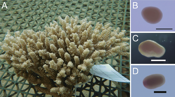
Fig. 1: Acropora tenuis (A) adult colony, (B) 3.5-day-old embryo, (C) 5.5-day-old planula, and (D) 36-day-old planula. Scale bars, 250 μm. Source: Kawamura et al., 2021.
In his study at Kochi University (Japan), Dr. Kawamura cultured A. tenuis in aquaria and induced spawning. After successful fertilization, embryos and planula larvae were cultured in the laboratory (Fig. 1). Cell dissociation was induced on planula larvae using both chemical (trypsin-EDTA-collagenase solution) and mechanical (rocking incubator) treatments. Cell lines grew in Dulbecco’s modified Eagle medium (DMEM), which is used for mammalian cell growth, diluted in seawater supplemented with HEPES (buffering agent) and antibiotics.
Dr. Kawamura and his colleagues achieved the dissociation of several cell types: brilliant, brown-coloured cells, translucent cells, and small, pale blue cells. These cell lines were able to create aggregates until week 2, then stopped growing but did not show signs of death. Furthermore, the application of a modular protease, plasmin, was effective in maintaining the dissociated cells in culture and in the appearance of new, dark cell lines. After 3 weeks, cell aggregates formed cell sheets and spheres resembling blastulas and gastrulas (two subsequent early stages of the embryotic development). However, blastula- and gastrula-like aggregates did not swim, nor developed into planula larvae. A total of eight representative cell lines (Fig. 2) were isolated during this experience. Cell lines were maintained for more than 8 months by replating them and were successfully cryo-preserved in liquid nitrogen.
Cultured cell lines
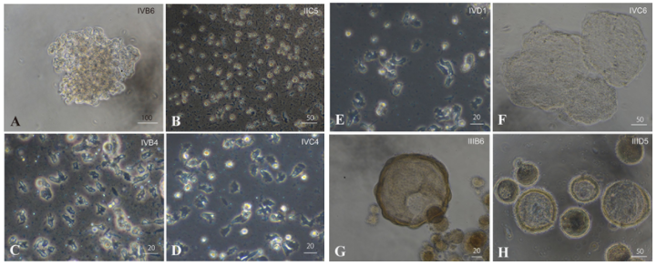
Fig. 2: Eight representative in vitro cell lines cultured by Dr. Kawamura. (A, B) Brilliant brown cell lines, (A) forming clusters, whereas (B) were stable single cells. (C-E) Flattened, dark, amorphous cell lines, (F) tend to form cell sheets. (H) Blastula-like cell clusters. (G) Gastrula-like cell clusters.
Bars: (A) 100 μm. (B, F, G) 50 μm (C-E and H) 20 μm. Source: Kawamura et al., 2021.
Immunochemical analysis
To ensure the cell lines did not result from contaminants, two tests were carried out. First, an immunochemistry analysis with coral-specific antibodies. Here, an antibody specific to a protein from A. tenuis was produced. Western blotting analysis was carried out against all proteins produced by planula larvae and cultured cell lines, confirming the specificity of the antibodies to A. tenuis. Afterwards, immunohistochemistry allowed the observation of ectoderm and endoderm stained with anti-antigens reacting with the protein (Fig. 3). Results confirmed that cultured cell lines belonged to A. tenuis.
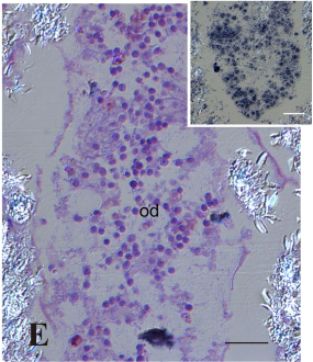
Fig 3: Anatomy of Acropora tenuis planula larvae. Section of the oral diverticulum stained with anti-antigens immunostaining. Note: cells are loosely associated. Source: Kawamura et al., 2021.
Molecular characterization
A transcriptomic approach was used to confirm the culture of A. tenuis cell lines and to characterize the genetic profiles of each cell line. The gene expression profile was determined using RNA sequence analysis on each cell type. Analysis used marker genes for gastroderm, gland/secretory cells, progenitors/undifferentiated cells, epidermis, neurons, larval neurons, larval apical organs, and dig filaments. All eight cell lines expressed 36 of 676 genes, the others being preferentially and/or specifically expressed in a certain line. This suggested that cell lines were not identical and did not have the same role in the development of the planula larvae.
Most of the expressed genes were categorized under “molecular function,”, followed by “biological function,” and “cellular component”. Gastroderm and glandular or secretory properties were particularly found in brilliant brown cells, suggesting their affinity to form living endoderm. Cell sheets were particularly rich in nervous system properties, and gastrula-like clusters likely revealed epidermis-like cell differentiation. Finally, amorphous cells and blastula-like cells had various cell functionalities, including gastroderm, glandular or secretory cells, and their progenitor cells.
Conclusions
Coral cell lines could reveal important evidence on the coral-symbiont relationship. Indeed, cell culture could offer an important device for the research in coral conservation, as well as in medicine, particularly in the study of stem cells and cell differentiation. Hence, assisting studies on reef sustainability and restoration, and on the mechanisms driving cell differentiation processes of planula larvae. Even though the technique for coral cell lines culture has recently been developed, using these cell lines could become key in the research on coral reefs and medicine.
Reference
Kawamura, K., Nishitsuji, K., Shoguchi, E., Fujiwara, S., & Satoh, N. (2021). Establishing Sustainable Cell Lines of a Coral, Acropora tenuis. Marine Biotechnology. https://doi.org/10.1007/s10126-021-10031-w
